NASH
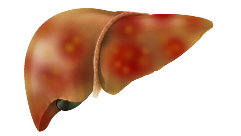
As the disease progresses, the liver undergoes scarring and fibrosis, which ultimately leads to cirrhosis. Fibrosis is characterized by the accumulation of a protein called collagen that can lead to increased liver stiffness. Liver fibrosis may ultimately lead to permanent organ damage, or progress to hepatocellular carcinoma or liver failure. |
 |
Biomarkers in NASH
Currently, diagnosis and staging of NASH is achieved through a liver biopsy, an invasive and potentially painful procedure. Biomarkers for liver fibrosis are under investigation to safely and rapidly diagnose the severity of disease and monitor changes in response to an intervention.
What is Magnetic Resonance Elastography (MRE)?
Magnetic Resonance Elastography (MRE) is a noninvasive, MRI-based imaging technique that measures tissue stiffness.
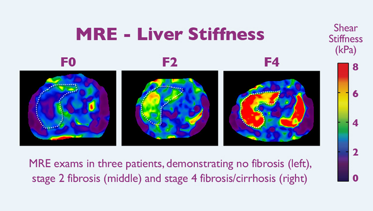
MRE uses MRI and mechanical vibrations to create color-coded cross-sectional images that display the stiffness of liver tissue. These images provide physicians a clear indication of the presence or absence of fibrosis in the entire liver,2 allowing radiologists to quantitatively and comprehensively measure changes in liver stiffness painlessly and noninvasively, as a quick and convenient add-on to the standard abdominal MRI procedure.

Research Implications
Liver stiffness, evaluated by MRE, is being used in clinical trials to:
- Provide a noninvasive biomarker to indicate the stage of liver fibrosis
- Measure changes in fibrosis while undergoing treatment
Liver stiffness, evaluated by MRE, is one of many biomarkers under investigation at Bristol Myers Squibb, and is being studied in collaboration with the Mayo Clinic.
Learn more about our work in fibrosis by visiting:
https://www.bms.com/researchers-and-partners/areas-of-focus.html